 Structure-based designs are integral to advancing drug discovery, and in this regard, cryogenic electron microscopy (cryo-EM) has emerged as a pivotal tool for achieving high-resolution structures of proteins and protein complexes. Particularly noteworthy is its utility in elucidating the structures of protein classes that have historically resisted crystallization.
Cryo-EM's pivotal role in structural biology was underscored by the joint awarding of the Nobel Prize in Chemistry in 2017 to Jacques Dubochet, Ph.D., Joachim Frank, Ph.D., and Richard Henderson, Ph.D., for their groundbreaking work in developing cryo-electron microscopy for the high-resolution structural analysis of biomolecules in solution[JS1] .
However, it's worth acknowledging that in the world of cryo-EM, the adage "garbage in, garbage out" remains just as relevant as in any other scientific endeavor. Cryo-EM's primary bottleneck, despite its apparent simplicity, lies in sample preparation. Biomolecules, it turns out, have a mind of their own – they can move, denature, aggregate, or arrange themselves in inconvenient orientations, all of which can severely impact the quality of the data obtained. Therefore, an improved sample grid design holds the potential to harness the capricious behavior of biomolecules.
Efforts are underway to democratize cryo-EM, a technology that has, at times, been prohibitively expensive. The pinnacle of cryo-EM instruments operates at 300 keV and carries a price tag of approximately $5-7 million. Nonetheless, there's progress on the horizon with the development of lower-cost instruments operating at around 100 keV, which come in at a more affordable $1-2 million. Among the pioneers in this arena are the scientists at the Structural Studies Division of the MRC [JS2] Laboratory of Molecular Biology at Cambridge University. Notably, Christopher J. Russo, PhD, who leads this division, recently authored a paper highlighting the potential advantages of 100 keV instruments. In the paper, it was noted that "Reducing the electron energy... offers both cost savings and potentially improved imaging," with lower energy electrons demonstrating a reduced propensity for radiation damage to biological specimens and an increased amount of information per unit damage.
Recent breakthroughs and ongoing challenges are essential to consider. Cryo-EM initially provided only indistinct images of biomolecules, limiting its application in structural biology. However, the last decade has witnessed significant advancements, with more efficient direct electron detectors, phase plates, and enhanced data processing software elevating the resolution to nearly atomic levels, typically around 3-4 Å, or even as low as 2.5 Å[JS3] . This has opened doors for structural insights into biological systems that are traditionally resistant to crystallization, such as membrane proteins. Nevertheless, it's worth noting that X-ray crystallography still boasts higher average resolution, albeit at the expense of introducing homogeneity.
One of the strengths of cryo-EM is its adaptability, with researchers consistently finding inventive ways to tackle complex scientific challenges. However, every cryo-EM endeavor must commence with meticulous sample preparation, which remains the foremost challenge in obtaining high quality data. Achieving this involves a complex and sample-specific process to generate a thin film of glassy ice containing randomly oriented intact molecules in biologically relevant conformations. Overcoming biomolecule movement during sample preparation and image capture is crucial, and this is accomplished through the use of grids coated with graphene and functional groups like amines, thiols, and carboxylic acids.
In recent times, functionalized grids and a plethora of other tools have emerged to aid cryo-EM practitioners, improving the ease, reproducibility, and quality of experiments. Companies like MiTeGen have played a vital role in engineering, manufacturing, and distributing products not only for cryo-EM but also for crystallization, crystal harvesting, cryocooling, and X-ray diffraction data collection across various biological and chemical compounds.
Addressing the challenges of manual cryo-EM sample preparation is a critical step forward. Traditional methods involve applying a few microliters of sample to a metal mesh, which is subsequently plunged into liquid ethane to vitrify the sample. This technique, while effective, carries significant variability, especially concerning blotting time and contact angle. Furthermore, the quality of a grid can only be ascertained once it is placed under the microscope.
In this regard, SPT Labtech has introduced the chameleon instrument, designed to automate the sample preparation process. It utilizes a camera-monitored robot to aspirate a small sample into a humidity zone, rotates the sample on a specially designed grid that eliminates the need for blotting, applies a hydrophilic charge to the grid, and employs an algorithm to ensure the desired liquid thickness. Subsequently, a robot plunges the grid into liquid ethane. This automated approach minimizes variability, with the time interval between sample application and freezing reduced to a remarkable 54 milliseconds – a vast improvement over traditional manual methods.
Moreover, cryo-EM's potential is further unlocked by employing faster plunge-freezing techniques, which mitigate challenges at the air-water interface such as preferred orientation or dissociation. Recent experiments, including those featuring the chameleon, indicate that increasing grid-making speed can help reduce particle damage and subunit dissociation.
The chameleon's rapid grid-making capabilities were instrumental in a study focused on the complex protein-folding process known as the GroEL-GroES chaperonin cycle. Faster plunge-freezing minimized denaturation at the air-water interface and improved particle orientation distribution, yielding a more detailed understanding of the chaperonin-assisted folding mechanism. These findings were presented in a recent preprint publication[JS4] , revealing that cryo-EM structures of stalled GroEL-ADP·AlF3-Rubisco-GroES complexes provided insights into Rubisco folding intermediates' interactions with GroEL-GroES via different sets of residues.
Another critical aspect highlighted by Thaw is the significance of the sample support in cryo-EM. Innovative developments like HexAuFoil, a gold hexagonal grid with a holey support film devised by Russo's group, promise to enhance data quality for a majority of samples.
Cryo-EM continues to expand its real-time data analysis capabilities. Edward Pryor, Ph.D, director of product management at Thermo Fisher Scientific, emphasizes the pivotal role of cryo-EM in advancing structural determination and drug discovery, particularly in debilitating neurodegenerative disorders and cancer. Thermo Fisher Scientific offers comprehensive cryogenic transmission electron microscopy (cryo.-TEM) solutions, including single-particle analysis, microcrystal electron diffraction, and cryo-electron tomography. Their portfolio includes cutting-edge instruments like the Tundra cryo-TEM, Glacios, and Krios cryo-TEMs, as well as cryo-FIB (focused ion beam) and cryo-PFIB (plasma focused ion beam) systems.
Pryor highlights that the Tundra, in particular, enables researchers to routinely achieve biologically relevant resolutions of 3-4 Å and prepare high quality samples for further analysis on Krios or Glacios cryo-TEMs. The ultrahigh-resolution Krios G4 cryo-TEM, configured for multiple applications, boasts the capability for rapid <2 Å resolution data acquisition, further elevating the potential for structural insights.
At Microscopy and Microanalysis 2022, Thermo Fisher Scientific partnered with Structura Biotechnology to introduce Embedded CryoSPARC Live, a software solution revolutionizing single-particle analysis in cryo-EM. This collaborative innovation promises to accelerate the transformation of raw data into high quality 3D protein structures, reducing the process from days to hours. Embedded CryoSPARC Live seamlessly integrates with Thermo Fisher's Smart EPU software and cryo-TEM technology, ushering in a new era of real-time cryo-EM data analysis.
While advancements in automation and software have streamlined the single-particle cryo-EM workflow, challenges persist, especially in data evaluation. Human expertise remains crucial in determining the suitability of acquired data for high-resolution structure determination.
Thermo Fisher's collaboration with Structura Biotechnology introduces a novel approach to single-particle cryo-EM data acquisition. By integrating Structura's CryoSPARC Live with Thermo Fisher's Smart EPU software and cryo-TEM technology, they enable real-time cryo-EM data analysis, making high-quality data accessible to users of all experience levels. This transformative approach significantly reduces the time required to convert raw data into high quality 3D protein structures.
In closing, the democratization of cryo-EM is an ongoing journey. The work of Russo and colleagues at the MRC Laboratory of Molecular Biology underscores the importance of reevaluating fundamental aspects of the technology. Their findings suggest that the optimal voltage for single-particle structural work is around 100 keV rather than the conventional 300 keV. This revelation led to the development of prototypes, including a dedicated modular field emission gun (FEG) operational at 100 keV. Such innovations pave the way for integrating this technology with existing electron columns, offering potential upgrades to thermionically operated TEMs and electron beam lithography systems for enhanced research and development applications. Cryo-EM's progress continues, driven by ingenuity and technological advancements that promise to unlock deeper insights into the structural biology of biomolecules and open new avenues for drug discovery.
1. Nobel Prize Outreach. The Nobel Prize in Chemistry 2017. Published October 4, 2017. Accessed August 2, 2023.
2. Hand E. Cheap shots. Science. 2020; 367(6476): 354–358. DOI: 10.1126/science.367.6476.354.
3. Naydenova K, McMullan G, Peet MJ, et al. CryoEM at 100 keV: a demonstration and prospects. IUCrJ. 2019; 6(Pt 6): 1086-1098. DOI: 10.1107/S2052252519012612.
4. Gardner S, Darrow MC, Lukyanova N, et al. Structural basis of substrate progression through the chaperonin cycle. bioRxiv. Posted May 31, 2023. DOI: 10.1101/2023.05.29.542693.
5. Klebl DP, Gravett MSC, Kontziampasis D, et al. Need for Speed: Examining Protein Behavior during CryoEM Grid Preparation at Different Timescales. Structure. 2020; 28(11): 1238–1248.e4. DOI: 10.1016/j.str.2020.07.018.
6. Naydenova K, Jia P, Russo CJ. Cryo-EM with sub-1 Å specimen movement. Science. 2020; 370(6513): 223–226. DOI: 10.1126/science.abb7927.
7. Peplow M. Cryo-Electron Microscopy Reaches Resolution Milestone. ACS Cent Sci. 2020; 6(8): 1274–1277. DOI: 10.1021/acscentsci.0c01048.
8. El-Gomati M, Wells T, Zha X, et al. A modular 100 keV vacuum sealed FEG for high-resolution electron microscopy. Microsc Microanal. 2021; 27(S1): 846–847. DOI: 10.1017/S1431927621003317.
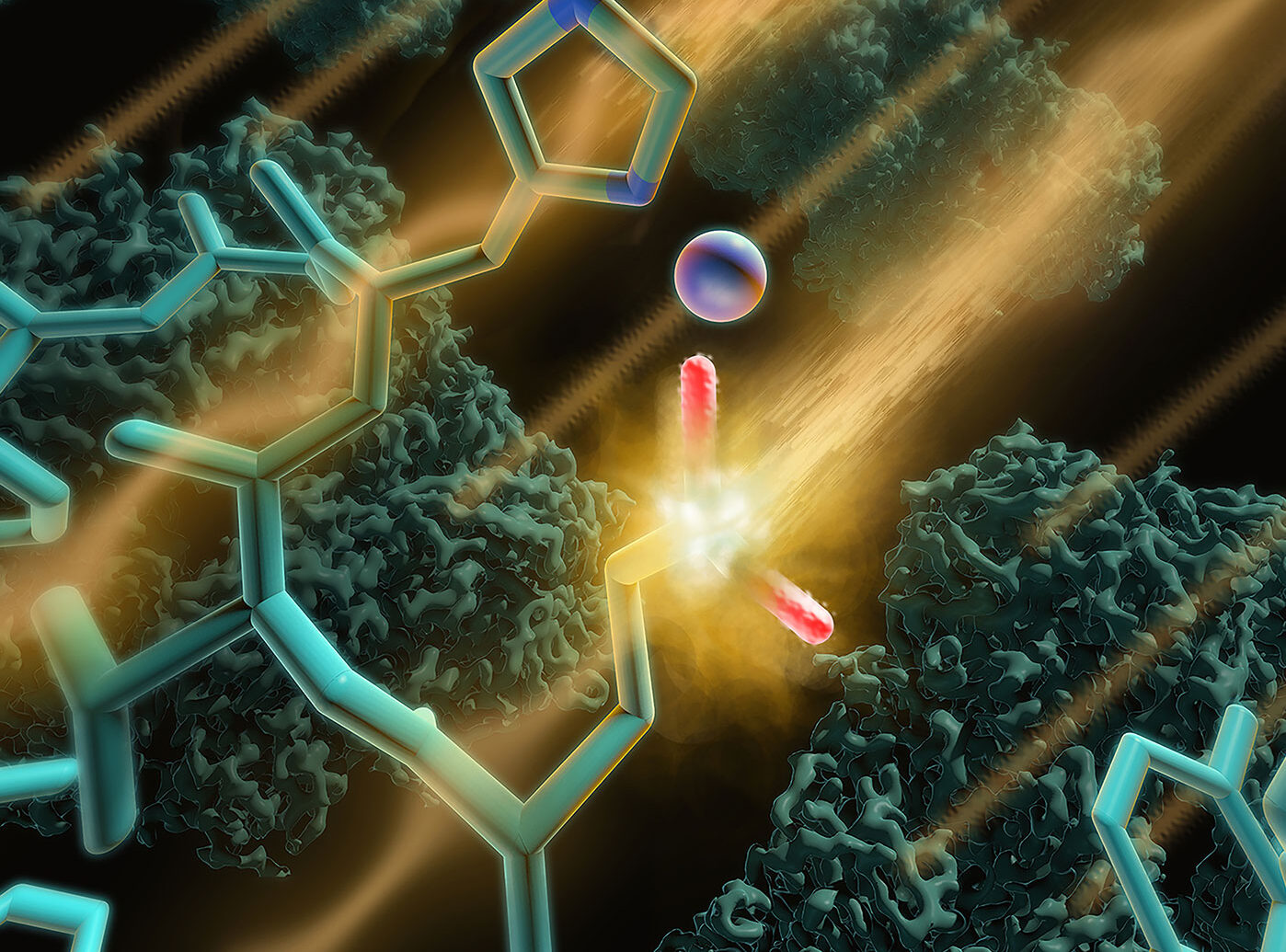
Image: The radiation emitted by high-resolution cryo-EM instruments can damage proteins. And some parts of proteins can be harmed more than others, complicating imaging. To reduce radiation damage and potentially improve imaging, investigators may opt to use less powerful instruments. Instruments rated at 100 keV are already being developed as less costly alternatives to 300 keV instruments. Credit: Veronica Falconieri, Sriram Subramaniam / NIH


