Beckman Institute Labs Bridge Advanced Imaging and Alzheimer’s Research
 The Beckman Institute for Advanced Science and Technology at the University of Illinois Urbana-Champaign has embarked on a life-changing research project to advance the early detection and diagnosis of Alzheimer’s disease. With the support of a $3 million grant from the US National Institute on Aging of the National Institutes of Health, the institute is leveraging the capabilities of its Magnetic Resonance Imaging (MRI) Laboratory and the Molecular Imaging Laboratory to develop innovative imaging techniques. Central to this initiative is the use of Cu-64 for Positron Emission Tomography (PET) imaging and Mn2+ for enhanced MRI, offering new possibilities for studying Alzheimer’s disease in living patients.
Alzheimer’s disease, a progressive neurodegenerative disorder, is characterized by the accumulation of amyloid plaques in the brain. Accurate diagnosis currently requires post-mortem analysis, underscoring the need for advanced diagnostic tools that can detect the disease at an early stage. The collaboration at Beckman aims to address this gap by combining the strengths of PET and MRI imaging, providing detailed insights into the physiological and pathological processes associated with Alzheimer’s.
The Beckman Institute’s utilization of cryogenic technology to support advanced biomedical research, particularly in the early detection and diagnosis of Alzheimer’s disease through sophisticated imaging methods, furthers the vital connection between cryogenics and biomedical imaging and highlights the wide-ranging applications and significance of cryogenic science in high-tech fields.
Cold Facts Invites You to Meet the Researchers
Two leading researchers at the Beckman Institute, Liviu M. Mirica and Wawrzyniec “Wawosz” Dobrucki, are at the forefront of pioneering research in Alzheimer’s disease. Their interdisciplinary project focuses on developing and applying novel imaging agents to revolutionize the diagnosis and treatment of this condition. Cold Facts sat down with these innovators to get a closer look at how their contribution is changing the conversation about a disease affecting so many.
Liviu M. Mirica (LMM) is a synthetic chemist, the William H. and Janet G. Lycan Professor of Chemistry in the School of Chemical Sciences and a researcher at the Beckman Institute. Mirica’s research group specializes in building and characterizing synthetic inorganic molecules in vitro, with a particular interest in developing novel radiopharmaceuticals for imaging and therapeutic purposes.
Wawrzyniec “Wawosz” Dobrucki (WLD) is a professor of bioengineering, the Neil and Carol Ruzic Scholar for Biomedical and Translational Sciences at the University of Illinois Urbana-Champaign and a researcher at the Beckman Institute. Dobrucki's research focuses on utilizing advanced imaging techniques to enhance our understanding of physiological and pathological processes.
Can you elaborate on the significance of using Cu-64 as the radioisotope for PET imaging in your newly acquired PET-CT scanner?
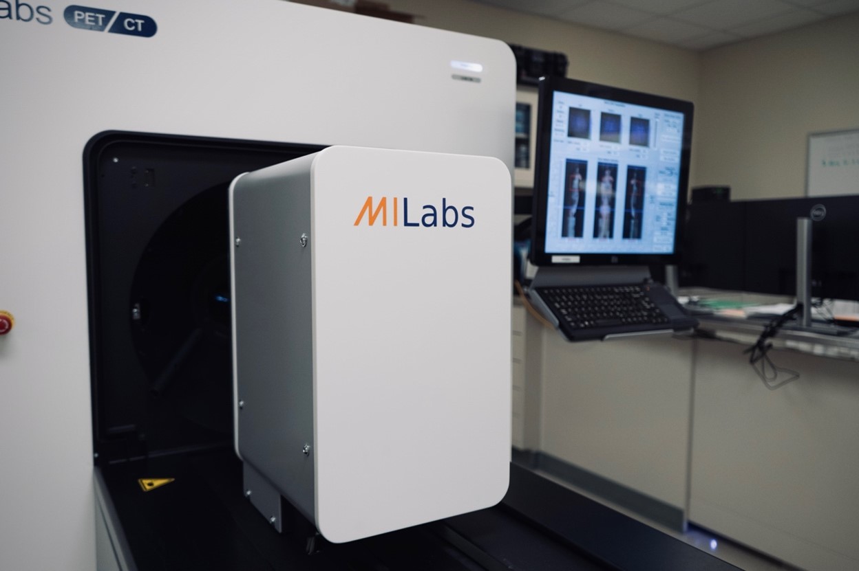
WLD: The energy of the positrons emitted by Cu-64 is suitable for high-resolution PET imaging. When used in conjunction with a high-performance PET-CT scanner like the U-PET7 from MILabs, Cu-64 can provide detailed images of physiological and pathological processes, improving diagnostic accuracy. In addition, Cu-64's properties make it suitable for quantitative imaging, which is crucial for accurately measuring biological parameters such as receptor density, blood flow and metabolic rates.
How does the use of Cu-64 enhance the capabilities of PET imaging compared to other radioisotopes commonly used in this modality?
LMM: Most clinical applications of PET imaging employ agents containing the C-11 and F-18 radiotracers, yet these radiotracers are limited by their short physical half-lives (20.4 min and 109.8 min, respectively). Therefore, the development of radio imaging agents containing longer-lived radionuclides would be advantageous as it would allow the possibility of imaging at longer time points where better contrast may exist. In addition, such PET agents can be shipped over longer distances and used in remote areas. In this regard, Cu-64 (t1/2 = 12.7 h) has become a useful radionuclide in the development of a wide variety of radiopharmaceuticals for imaging and therapeutic purposes.
What specific biological processes or conditions do you anticipate studying with the aid of Cu-64 PET imaging?
LMM: We are developing better diagnostic tools and imaging agents for the early detection of Alzheimer’s disease. A key marker of Alzheimer’s disease is the presence of amyloid plaques, or aggregates, in the patient’s brain. Currently, diagnosing Alzheimer’s disease accurately can only be completed during post-mortem investigation. Thus, the development of new imaging agents as diagnostic tools that can rapidly assess the presence of early-stage amyloid aggregates in a living patient can have a large health-related impact.
Moving to MRI imaging, why have you chosen Mn2+ as the paramagnetic ion to provide improved contrast?
LMM: While PET imaging is by far the most used imaging modality in Alzheimer’s disease, it can be invasive and expensive. Magnetic resonance imaging (MRI) has emerged as a potential complement to PET analysis, since it can also provide high-resolution images of the brain that would be beneficial for early diagnosis. In order to improve the quality of MR images, contrast agents based on paramagnetic ions such as gadolinium (Gd) are used to alter the magnetic properties of nearby water molecules; however, these Gd agents have drawbacks given their accumulation in the body and toxicity. Thus, the development of MRI contrast agents based on other less toxic paramagnetic ions such as Mn2+ would be extremely useful for early Alzheimer’s
diagnosis.
How do you foresee the combination of PET imaging with Cu-64 and MRI imaging with Mn2+ contributing to interdisciplinary research projects at your institution?
LMM: This joint research project is one of the first federally funded grants to bridge upcoming research between Beckman’s Magnetic Resonance Imaging Laboratory and the Molecular Imaging Laboratory.
Are there any particular research areas or clinical applications where the synergistic use of these imaging modalities holds particular promise?
LMM: By developing multimodal imaging agents, we hope to employ the advantages of each imaging modality, while reducing their disadvantages. On one hand, PET imaging is highly sensitive but requires the use of a radiotracer that needs to be injected into a patient. On the other hand, MRI is an imaging technique that can produce high-resolution images but requires contrast agents to create the desired high contrast.
Could you discuss any challenges or considerations involved in integrating Cu-64 PET imaging and Mn2+-enhanced MRI into your research protocols?
LMM: The main challenge we are trying to address is the unprecedented development of chelating agents that can target the amyloid aggregates found in the brains of Alzheimer’s patients and can be labeled separately with Cu-64 and Mn2+ for PET and MRI diagnostic applications, respectively.
In terms of practical implementation, what steps are being taken to ensure the safe and effective utilization of Cu-64 and Mn2+ in imaging studies?
LMM: Since Cu-64 is a radiotracer, stringent safety protocols need to be put in place where PET imaging studies are being performed. Fortunately, at the Beckman Institute both the MRI lab and the Molecular Imaging Lab for PET imaging are close to each other in the basement of the institute.
Looking ahead, do you envision any future advancements or developments in the use of these imaging agents for even greater insights into biological processes or clinical diagnoses?
LMM: The development of multimodal PET and MRI diagnostic agents for Alzheimer’s diseases could then be extended to other neurodegenerative disorders such as Parkinson’s, Lewy Body dementia (LBD) and Frontotemporal degeneration (FTD), or for broad neuroimaging applications.
Image: This PET machine located in Beckman's Molecular Imaging Laboratory will be operated by Dobrucki and used extensively during the team's research. Credit: Beckman Institute for Advanced Science and Technology at the the University of Illinois Urbana-Champaign


